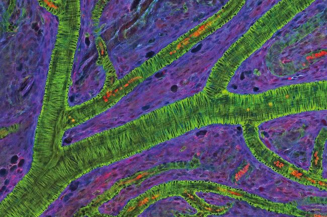
Behind your eyes
© Alamy
This is an image of the blood vessels at the back of the eye, the retina, under a microscope. The green branch-like structures indicate the size and shape of blood vessels. The stain used in this microscopy technique adheres to filaments of proteins, called actin, that wrap around each blood vessel. Although the majority of blood cells were removed to produce this image, some remain, and can be seen as red flecks.